

TOF Technology in Biological Experiments: 3D Observation & Precision
How can TOF technology be used in biological experiments to achieve 3D microscopic observation and high-precision data acquisition?
With the continuous development of life sciences, researchers increasingly require three-dimensional observation of cellular dynamics, tissue structures, and microscopic processes. However, traditional microscopy has clear limitations in dynamic 3D imaging and real-time data acquisition. TOF (Time-of-Flight) (https://en.wikipedia.org) microscopy leverages its depth sensing capabilities and high-speed imaging advantages to provide a new research tool for biological experiments, driving studies toward high precision, visualization, and intelligent analysis.
What is Electron Microscopy Used For?
Electron microscopes (EM) in biological experiments and microscopy are mainly used for:
-
High-Resolution Structural Observation
Electron microscopes use electron beams to image samples, revealing nanoscale structures of cells, tissues, or materials far beyond the resolution of traditional optical microscopes. -
Subcellular and Microscopic Tissue Analysis
In studies of cell migration, neuronal outgrowth, or angiogenesis, EM provides detailed images of organelles, membranes, and microstructures, offering reliable references for experimental data. -
Multi-Modal Experiment Integration
EM is often combined with TOF microscopy to achieve complementary 3D dynamic observation and ultra-high-resolution structural analysis. For example, TOF provides real-time 3D spatial information, while EM delivers nanoscale detail, allowing precise monitoring of cell movements, tissue engineering, or microscopic drug screening processes. -
Data Visualization and Quantitative Analysis
Combined with TOF 3D point clouds or AI algorithms, high-resolution EM images can be used for quantitative analysis of cell volume, morphology, and spatial distribution, assisting researchers in high-precision experimental studies.
In summary, electron microscopy is primarily used for high-resolution observation and microstructure analysis. It is especially suitable for integration with TOF microscopy to achieve dynamic 3D observation and nanoscale detail analysis, providing reliable data and visualization support for biological experiments, tissue engineering, and microscopic drug screening.

The Need for 3D Observation in Biological Experiments
In advanced fields such as cell biology, tissue engineering, and microscopic drug screening, scientists increasingly focus on the dynamic 3D behaviors of microscopic objects. For instance, cultured cells not only migrate but also extend, divide, and aggregate to form complex spatial networks; in tissue engineering, cells grow within 3D scaffolds, constructing tissue structures that mimic the in vivo environment; in drug screening experiments, drug effects on cells or tissues often involve spatial and temporal changes. Observing and quantifying these processes is critical for understanding biological mechanisms, optimizing experimental design, and improving reproducibility.
Traditional optical microscopes and 2D imaging techniques have obvious limitations in these experiments. 2D microscopes provide only planar images, failing to fully reflect cell movement trajectories, tissue spatial distribution, and microstructural changes, particularly in complex 3D environments. For example, during tumor cell migration studies, 2D images only provide planar projections and cannot accurately quantify cell speed, direction, or morphology in 3D space. In neuroscience, 2D imaging cannot fully capture the 3D growth paths and branching patterns of neurons. In angiogenesis experiments, 2D imaging cannot precisely measure the 3D topology and branch angles of vascular networks.
Additionally, 2D observation can introduce errors and biases. Researchers often rely on continuous slicing or multi-angle imaging to infer 3D structures, increasing experimental time and complexity while potentially affecting the accuracy and scientific validity of data. This limitation is especially pronounced in high-throughput experiments or live-cell dynamic observation.
Thus, high-precision, real-time 3D dynamic observation has become an urgent requirement in modern life science experiments. TOF (Time-of-Flight) microscopy emerged in this context. By measuring the time it takes for light pulses to travel to and reflect back from a sample, TOF rapidly generates 3D spatial data, achieving non-contact, real-time, dynamic microscopic 3D observation. With TOF, researchers can quantify cell migration trajectories, monitor cell distribution in tissue engineering, and perform high-precision analysis of microscopic drug effects, greatly improving experimental reproducibility, accuracy, and scientific value.
In short, as biological research becomes more complex and refined, traditional 2D microscopy can no longer meet scientific needs. TOF microscopy’s 3D dynamic observation capability provides a new technological foundation for microscopic life science research, advancing experiments toward intelligent, high-throughput, and quantifiable directions.
TOF Microscopy Principles
TOF (Time-of-Flight) microscopy is a high-precision 3D imaging technique based on light pulse time-of-flight measurement. Its core principle involves emitting short light pulses (usually infrared) toward a target sample and precisely measuring the time difference between emission and the reflected return to the sensor, thereby calculating the spatial depth information of microscopic structures. In other words, TOF converts the “flight time” of light directly into 'spatial distance,' generating complete 3D depth data.
1. Technical Advantages
-
3D Depth Perception
TOF generates complete 3D spatial data of microscopic samples, allowing researchers to observe cell morphology, tissue layers, and dynamic microstructural changes. This is essential for understanding cell migration paths, cell distribution in tissue scaffolds, and the spatial relationships of complex microenvironments. -
High-Speed Imaging and Real-Time Observation
TOF supports millisecond-level imaging, capturing dynamic biological processes such as membrane motion, neuronal outgrowth, angiogenesis, and cell growth in tissue engineering, providing continuous high-frequency data streams for experiments. -
Non-Contact Measurement
TOF requires no direct contact with the sample, avoiding interference from dyes, labels, or probes. It is ideal for live cells, sensitive tissues, or fine microstructures and reduces sample damage, improving experimental safety and data reliability.
2. TOF vs. Electron Microscopy (EM)
In microscopic imaging, electron microscopes (EM) are another widely used tool. EM uses electron beams instead of light to illuminate samples and leverages electron wave properties to achieve resolutions far beyond optical microscopes. EM is commonly used for:
-
Organelle and microstructure analysis (mitochondria, ribosomes, membrane structures)
-
Materials science (nanostructures, crystal defects)
-
Virus and microorganism studies (fine viral particles or microbial structures)
The working principle of EM includes electron beam emission, sample-electron interaction, electron detection, and signal conversion into high-resolution images. EM can reveal nanometer- or even sub-nanometer-scale structures but usually requires sample preparation (fixation, dehydration, embedding, staining) and cannot easily perform real-time dynamic observation.
In comparison, TOF microscopy, while slightly lower in resolution than high-end EM, excels in 3D dynamic observation, non-contact measurement, and real-time imaging, making it particularly suitable for continuous monitoring in biological experiments and live sample research.
3. Applications of TOF in Biological Experiments
-
Cell Migration and Morphology Analysis: Real-time capture of cell movement trajectories and shape changes.
-
Tissue Engineering: Observation of cell distribution and tissue formation within 3D scaffolds.
-
High-Throughput Drug Screening: Non-contact monitoring of drug effects on cell growth and behavior.
-
Microstructural Quantitative Analysis: Integration with AI algorithms for visualization and quantitative assessment of depth data.
4. Related Measurement Tools and Derivative Technologies
In addition to TOF and electron microscopy, modern laboratories may use the following technologies to supplement experimental data:
-
Electronic Distance Meter (EDM): Precise measurement of micro- or macroscopic distances.
-
Capacitive Humidity Sensor: Monitoring experimental environment humidity to provide stable conditions for sensitive samples.
In conclusion, TOF microscopy, with its non-contact, high-speed, 3D dynamic observation capabilities, provides powerful data support for modern life science experiments. Combined with traditional electron microscopy, it offers complementary advantages, enhancing experimental efficiency and precision while enabling high-throughput, dynamic monitoring, and intelligent data analysis.

Experimental Cases and Research Achievements
TOF (Time-of-Flight) technology has shown immense potential in biological experiments, providing unprecedented 3D dynamic observation and quantitative analysis capabilities for life science research. Its applications include the following areas:
1. Cell Migration Analysis
In oncology, immunology, and stem cell research, cell migration is a key research indicator. Using high-precision 3D point cloud data generated by TOF, researchers can track the trajectories of individual or groups of cells in real time, including migration speed, direction, and morphological changes. For example, in tumor metastasis studies, TOF can accurately quantify cancer cell movement through the extracellular matrix, providing reliable data for understanding metastatic mechanisms. In immune cell studies, TOF captures rapid morphological changes of cells in microenvironments, helping assess immune response efficiency.
2. Tissue Engineering and 3D Scaffold Monitoring
In tissue engineering and artificial organ cultivation, monitoring cell seeding and tissue remodeling within 3D scaffolds is critical. TOF microscopy can track cell distribution, proliferation, and morphological changes in scaffolds in real time, recording tissue growth dynamics and providing data to optimize culture conditions. For instance, in liver or cardiac organoid construction experiments, TOF can observe the formation of tissue layers, identify abnormal growth or structural defects, and support quality control and process improvement.
3. Data Visualization and Quantitative Analysis
TOF-generated 3D data are not only intuitive but can also be combined with AI and deep learning algorithms for automated quantitative analysis. Researchers can count cells, measure volume, assess morphology, and even track tissue dynamics. For example, combining TOF with machine learning allows automatic plotting of tumor growth curves, dynamic tracking of neuronal outgrowth, or quantification of angiogenesis rates. This automated processing significantly improves experimental efficiency, reduces human error, and enhances reproducibility.
4. Support for Live and Microenvironment Experiments
TOF’s non-contact measurement advantage makes it suitable for live cell and microenvironment experiments, providing high-precision data without staining or labeling. This is particularly important for sensitive tissues or long-term cultures, preserving sample integrity while capturing continuous dynamic changes, offering strong technical support for drug screening, stem cell research, and microenvironment regulation experiments.
These experimental cases demonstrate that TOF technology provides high-precision 3D data and real-time dynamic observation, along with data quantification and automated analysis, greatly enhancing accuracy, efficiency, and reproducibility. Whether for cell migration studies, tissue engineering, drug screening, or microenvironment analysis, TOF microscopy shows strong potential as a core tool for modern life science experiments.
Technical Challenges and Optimization Directions
Although TOF biological imaging shows great promise in microscopy and 3D observation experiments, it still faces several technical challenges in practical applications. Researchers conducting cell migration, tissue engineering, or microscopic drug screening studies need to address the following issues:
1. Technical Challenges
-
Resolution and Sensitivity Limitations
Although TOF provides 3D depth information, at the microscopic scale, light signals can attenuate or scatter, making fine structures difficult to resolve. This is particularly critical for live cells or sensitive tissues. -
Dynamic Sample Interference
Cell motion, membrane activity, and tissue growth can produce small dynamic changes that affect TOF imaging stability and accuracy. -
Complex Experimental Environment Interference
Changes in laboratory lighting or interference from electronic instruments (such as electronic distance meter definition and capacitive humidity sensor) may introduce noise into TOF signals. -
Compatibility with Traditional Microscopy
Researchers often combine electron microscope or electron microscopy for high-resolution observation, but achieving complementary data integration between TOF and electron microscopy (electron microscop/electron microcopy) remains a technical challenge.
2. Optimization Directions
-
Light Source and Wavelength Optimization
Selecting light pulse wavelengths suitable for microscopic samples improves penetration and reflection stability, enhancing imaging precision of microstructures. -
Advanced Signal Processing Algorithms
AI and deep learning can denoise, fuse, and perform multi-frame accumulation on TOF point cloud data, achieving real-time 3D reconstruction of dynamic samples, similar to how electron microscopes work in electron microscopy. -
Multi-Modal Microscopy Integration
Combining TOF with conventional optical microscopes and electron microscopes (electron microscopes/electron microscope/electronic microscope) creates complementary imaging systems. TOF provides high-real-time 3D information, while electron microscopy provides ultra-high-resolution structural detail, significantly improving experimental reliability. -
Hardware Upgrades
Developing high-sensitivity, low-noise TOF sensors, optimizing light source power and receiver performance, and improving spatial resolution and field of view ensure precise and stable data in complex environments. -
Dynamic Environment Adaptation
Real-time depth tracking and adaptive calibration algorithms allow TOF systems to handle sample movement and environmental changes, ensuring continuous, reliable 3D observation.
Through these optimizations, TOF biological imaging can overcome limitations in resolution, scattering, and dynamic interference, and integrate with traditional microscopy and intelligent algorithms to provide more accurate, stable, and efficient experimental data support.

Future Development Trends
With the rapid development of TOF microscopy, AI, and intelligent experimental equipment, biological research is transitioning toward automation, high-throughput experiments, and 3D dynamic visualization. Future trends of TOF applications in research include:
1. TOF + AI for Automated Experiments
Combining AI algorithms, TOF point cloud data can automatically identify cells, tissue structures, and dynamic changes:
-
Automatic Annotation and Analysis: Systems can automatically identify cell migration trajectories and tissue growth patterns, improving efficiency and reproducibility.
-
Precise 3D Measurement: Generate 3D models of microstructures to quantify volume, morphology, and spatial relationships.
-
High-Throughput Screening: Support large-scale experiments such as drug screening or gene editing, reducing manual operation and error.
2. Multi-Modal Experiments with Electron Microscopy
TOF can be combined with electron microscopy or electron microscopes (electron microscope/electron microscopes/electronic microscope) for complementary data:
-
3D Observation and Ultra-High-Resolution Fusion: TOF provides dynamic 3D spatial information, electron microscopy delivers nanoscale details.
-
Precise Experimental Planning: Researchers can design observation schemes in 3D simulations before experiments, optimizing microscopy workflows.
-
Cross-Scale Research: Achieve continuous observation and analysis from cellular to subcellular levels.
3. High-Precision Experimental Instrument Development
-
Integrated Sensors: Combine TOF with capacitive humidity sensors and electronic distance meters to achieve environment-adaptive adjustments.
-
High-Speed Imaging: Millisecond-level scanning meets dynamic process recording requirements and supports real-time analysis and feedback.
-
Safe, Non-Invasive Observation: Non-contact measurement protects live cells and sensitive tissues.
4. Intelligent Laboratory Construction
Integrating TOF with AI, cloud platforms, and automated instruments promotes smart laboratory development:
-
Remote Monitoring and Experiment Management: Researchers can access TOF data remotely for guidance and analysis.
-
Data Visualization and Report Generation: 3D point cloud data combined with AI can automatically generate experimental reports and statistical analysis.
-
High Throughput and Reproducibility: Intelligent systems reduce human error, improve efficiency, and enhance data reliability.
Conclusion
TOF microscopy, with its 3D observation, non-contact measurement, and high-speed imaging advantages, is transforming biological research. From cell migration analysis and tissue engineering monitoring to high-throughput drug screening, TOF not only improves experimental precision but also optimizes research workflows. Combined with AI, multi-modal microscopy (such as electron microscopy), and intelligent experimental platforms, TOF will become a core technology in future laboratories, providing more precise, efficient, and intelligent solutions for life science research.
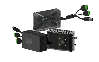
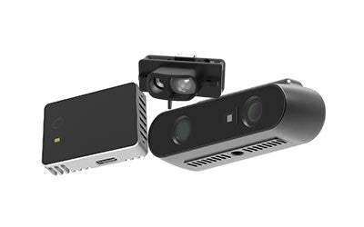
Synexens Industrial Outdoor 4m TOF Sensor Depth 3D Camera Rangefinder_CS40p

After-sales Support:
Our professional technical team specializing in 3D camera ranging is ready to assist you at any time. Whether you encounter any issues with your TOF camera after purchase or need clarification on TOF technology, feel free to contact us anytime. We are committed to providing high-quality technical after-sales service and user experience, ensuring your peace of mind in both shopping and using our products.