

TOF Medical Imaging : 3D Diagnosis for Minimally Invasive Surgery
How can TOF technology be used to achieve precise diagnosis and 3D medical imaging in minimally invasive surgeries?
With the advancement of modern medical technology, precise diagnosis and minimally invasive treatment have become key trends in healthcare. However, traditional medical imaging techniques still face limitations, failing to fully meet clinical demands for three-dimensional spatial information and real-time monitoring. TOF (Time-of-Flight) (https://en.wikipedia.org)medical imaging, with its high-precision depth sensing capabilities, offers innovative solutions for minimally invasive diagnosis and medical imaging. It is gradually becoming a core technology for surgical planning, lesion localization, and intelligent diagnostics.
What is TOF Time-of-Flight Technology?
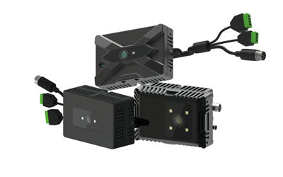
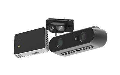
TOF (Time-of-Flight) technology is a depth sensing method based on measuring the travel time of light pulses. It works by emitting light pulses (usually infrared) toward a target and measuring the time difference from emission to reflection back to the sensor. This allows precise calculation of the distance between the sensor and the object.
In the context of medical imaging, rehabilitation, and health monitoring, TOF technology has the following characteristics:
-
3D Depth Perception: Generates 3D spatial data of organs, lesions, or the human body, supporting organ volume measurement, lesion localization, motion capture, and posture analysis.
-
Non-contact Measurement: No need for wearable devices or direct contact, enabling non-invasive or minimally invasive procedures.
-
High Real-Time Performance: Captures data in milliseconds, supporting fast response and real-time monitoring, such as surgical navigation, rehabilitation feedback, and fall detection.
-
Multi-Scenario Applicability: Used in medical imaging, minimally invasive diagnosis, rehabilitation training, elderly health monitoring, and smart home health management.
In short, TOF technology converts time measurements into spatial depth information, enabling precise, real-time, and three-dimensional sensing and monitoring.

Limitations of Traditional Medical Imaging
Traditional medical imaging faces limitations not only in image dimensions but also in clinical safety and efficiency. While X-rays, CT, and 2D ultrasound are widely used, they have clear constraints in complex surgeries or minimally invasive procedures.
1. Lack of Spatial Information
2D imaging provides only planar projections and cannot accurately reflect the three-dimensional shape and spatial relationship of organs. In tumor localization or vascular branch assessment, 2D images cannot visually present the precise location of lesions, increasing the risk of errors during surgery and potential damage to organs, nerves, or blood vessels.
2. Surgical Planning Errors
Minimally invasive surgeries have limited operating space and restricted vision. Preoperative planning based on 2D images often relies on experience to estimate spatial relationships between instruments and tissues. Misestimations can lead to deviations in surgical access and operating angles, compromising safety and success, especially in complex anatomical areas like the brain, heart, or deep abdominal tissues.
3. Limited Diagnostic Efficiency
2D imaging requires multiple slices or projections for analysis, which increases diagnostic time and relies heavily on the physician’s spatial reasoning. For less experienced doctors, this may increase the likelihood of misjudgment and affect clinical decisions.
4. Challenges for Minimally Invasive and Complex Surgeries
Minimally invasive surgeries demand precise navigation and instrument control. Traditional 2D imaging cannot provide real-time depth information or track dynamic changes in tissues, limiting surgical precision, especially in laparoscopic, arthroscopic, or interventional procedures.
In summary, while traditional imaging remains essential, its 2D limitation, planning inaccuracies, and inefficiencies necessitate 3D imaging and depth sensing technologies to enhance precision, efficiency, and safety in minimally invasive and complex procedures.
TOF for 3D Medical Imaging
TOF enables 3D medical imaging, overcoming the limitations of 2D imaging and providing clinicians with precise, intuitive 3D data for surgical planning, diagnostic analysis, and intraoperative navigation, thereby improving safety and efficiency.
1. Real-Time Depth Scanning
TOF systems emit light pulses or infrared signals and measure reflection times to capture depth information within milliseconds. This rapid imaging allows surgeons to visualize organ structures and lesion positions in real time, without waiting for CT or MRI results, improving intraoperative responsiveness.
2. Accurate Organ and Lesion Volume Measurement
2D imaging cannot precisely calculate organ or lesion volumes. TOF 3D imaging generates full point cloud data for spatial reconstruction, enabling accurate volume and shape assessment. Surgeons can determine resection ranges in tumor operations, minimizing damage to surrounding healthy tissue.
3. Precise Lesion Localization
TOF provides 3D spatial coordinates for lesions, tumors, or blood vessels, enabling surgeons to plan minimally invasive paths accurately and avoid critical structures, increasing intraoperative control and safety.
4. Dynamic Tissue Tracking and Real-Time Navigation
Organs move subtly due to respiration, heartbeat, or minor patient motion. TOF’s high refresh rate and real-time depth sensing capture these changes. When combined with surgical robots or navigation systems, surgeons can adjust procedures dynamically for enhanced precision.
5. Assisted Diagnosis and Personalized Treatment
TOF 3D imaging generates intuitive visual reports, evaluating lesion shape, location, and surrounding tissue relationships. Combined with AI, TOF supports lesion recognition, risk prediction, and personalized treatment planning, advancing precision medicine.
Compared to traditional 2D imaging, TOF provides superior spatial visualization, real-time feedback, dynamic tracking, and volumetric measurement, making it essential for minimally invasive surgery, complex procedures, and personalized treatment.

Clinical Applications
TOF shows broad clinical potential, particularly in minimally invasive diagnosis, surgical navigation, and precision treatment. Its advantages include real-time 3D depth acquisition, dynamic tissue tracking, and quantifiable, intuitive data to improve surgical accuracy and safety.
1. Minimally Invasive Surgical Navigation
TOF provides 3D depth images for precise surgical path planning:
-
Minimized Incisions: Surgeons can select the smallest access path, reducing tissue damage and recovery time.
-
High-Precision Operations: Essential in neurosurgery, cardiac, and laparoscopic procedures to identify critical structures and reduce risks.
-
Dynamic Strategy Adjustment: TOF tracks organ motion (e.g., respiration, heartbeat), enabling real-time surgical adjustments.
2. Tumor Localization and Volume Measurement
Accurate tumor positioning and volumetric assessment support resection, radiotherapy, and targeted treatment:
-
3D Localization: TOF provides precise 3D tumor coordinates for surgical or treatment planning.
-
Dynamic Volume Monitoring: Multiple scans allow real-time tumor monitoring, guiding treatment optimization.
-
Targeted Therapy: Combined with AI, TOF assists precise drug or energy delivery to lesions, sparing healthy tissue.
3. Vascular Imaging and Interventional Therapy
TOF offers reliable 3D guidance in vascular interventions:
-
3D Vascular Reconstruction: Aids catheter or stent navigation.
-
Reduced Complications: Precise data lowers risk of vessel injury or misplacement.
-
Intraoperative Monitoring: Continuous tracking of vessels and surrounding tissues provides actionable guidance.
4. Personalized Treatment and Rehabilitation Support
TOF supports preoperative planning and postoperative recovery:
-
Tailored Surgical Plans: 3D models of patient anatomy enable customized surgery.
-
Rehabilitation Tracking: Monitors joint range of motion and posture post-surgery, informing rehab strategies.
-
Remote Medical Assistance: Cloud-based data and AI enable remote evaluation, intraoperative guidance, and rehab management.
In conclusion, TOF medical imaging enhances diagnostic precision, optimizes minimally invasive procedures, and improves dynamic monitoring, providing a highly efficient, safe, and visualized tool for modern healthcare. With further integration with AI, surgical robotics, and remote medical platforms, TOF’s clinical applications will continue to expand and become increasingly intelligent.

